The magnifying power of clear glass and crystal was probably discovered by the early Greeks and Babylonians. Can you imagine the amazement of an artisan who, looking through a primitive lens, is suddenly exposed to the ferocity of weevils infesting their bread, or the wondrous detail in a feather or cobweb? Would we have inherited the brilliance of Galileo and Copernicus, several hundred years later, if not for these early crafts-folk?
A glass lens will bring distant objects closer, make the smallest particles appear larger, and provide relief for our aging eyes as our natural lenses harden with age (a condition called presbyopia – spectacles were probably invented around the 11th century). Even the simplest microscopes and telescopes will expand your universe.
Macrofossils, the kind we easily see, were the primary instruments used in the 18th and 19th centuries to establish an ordering of strata, or stratigraphy. William Smith’s monumental goal to create a geological map of England would have been next to impossible without a macrofossil-based stratigraphy. Microfossils, the kind we need a microscope to see, have added a huge level of detail and sophistication to our stratigraphic studies, such that sequences of rock can be finely tuned with respect to their relative geological age. For example, the macro- and microfossils in strata at one locality can be compared with fossil assemblages in strata at a more distant location to determine whether they are the same age, and whether the ancient environment in which they lived was similar.
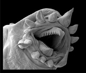
Our understanding of microfossils and the role they play in stratigraphy, has flourished in concert with the technical advances in microscopes; from the simple uni-ocular, to the binocular, and beginning in the 1960s, (the first commercial) scanning electron microscopes (SEMs). In fact, SEMs were like a paradigm shift in visualizing microscopic critters.
Most sedimentary rocks contain microfossils of one kind or another. They are present in marine deposits and terrestrial deposits such as rivers, lakes, and soils, which means they can be used to compare, by age or environment, ancient deposits from a diverse set of environmental conditions. There are many kinds of microscopic life forms and fragments of life forms, but the groups paleontologists most commonly use include:
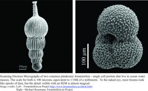
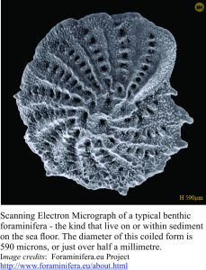
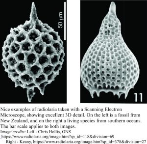
- Foraminifera (single cell protists) and Radiolaria that range from the shallowest marine settings (like tidal flats and estuaries) to the deepest oceans (marine deposits only). Both produce hard shells or skeletons, and in the case of Forams (as they are commonly called) the shells are made of calcium carbonate, whereas radiolaria skeletal material is made of silica (like quartz).
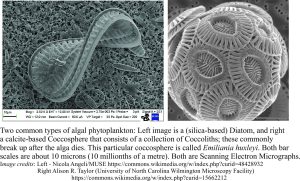
- Various groups of algae such as Diatoms (with skeletons of silica, from which diatomaceous earth derives), that can live in both marine and freshwater environments, and Coccoliths that are predominantly marine phytoplankton and secrete calcium carbonate skeletons; they are one of the main constituents in natural chalk (like the White Cliffs of Dover). Coccoliths are one of the culprits responsible for marine algal blooms.
- Pollen and spores from plants – these are common in river, lake and swamp-bog deposits, but pollen can also be transported by wind and accumulate with marine microfossils on the sea floor. These fossils consist of organic compounds.
One of the acknowledged problems in stratigraphy is deciphering the relationship between deposits formed in marine and non-marine (i.e. land-based fresh water) environments. This problem exists because most animal and plant species tend to live in one or the other; they either live in the sea, or on land or fresh water (some specialized plants like Mangroves, straddle both environments). Fossil pollen and spores help bridge this gap, partly because they are so mobile; pollen can be transported by wind many 100s of kilometres from its source.
All these fossil groups have living relatives, so that people who study such things can compare the modern forms with their more ancient representatives. However, there are some microfossil groups that are extinct. Conodonts are not complete animals in themselves (like Forams) but are tooth-like fragments of an extinct eel-like animal group that lived from the early Cambrian Period (542 million years ago), to the Triassic Period, about 200 million years ago. Conodonts are a very important fossil group because they permit refined age determination of Paleozoic rocks; they may also be some of the earliest, ancestral vertebrates.
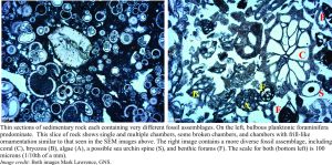
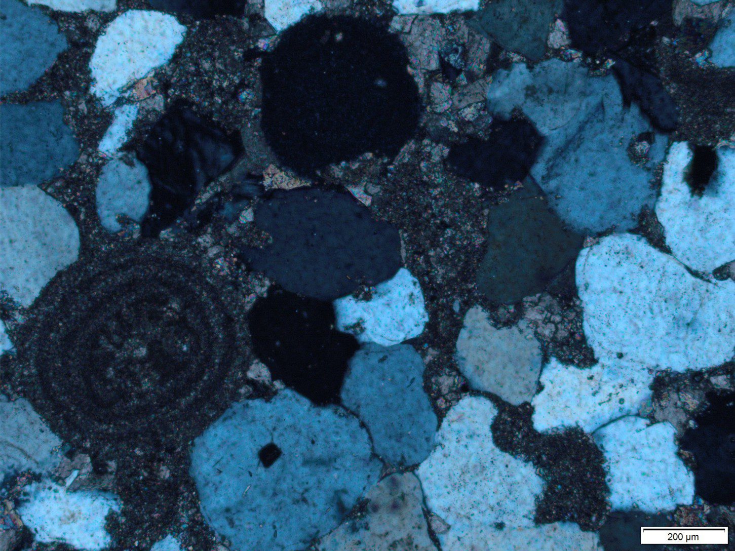
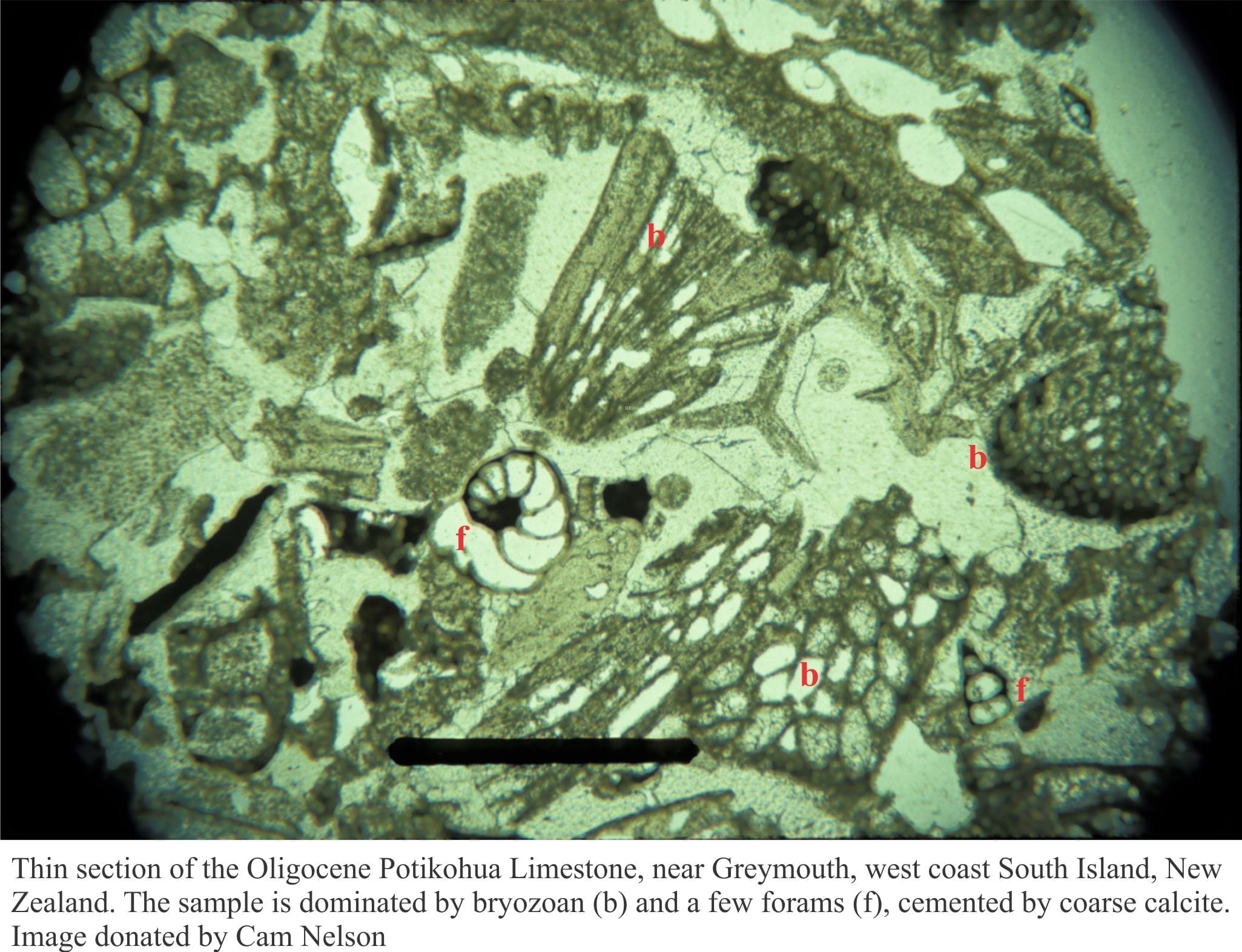
All the microfossil images depicted so far, are SEM photos that present in extraordinary detail the three-dimensionality of skeletal and shell structure, and ornamentation. However, the history of rock formation, and its journey from deposition to burial, uplift and erosion, are better illustrated in thin slivers of rock, or thin sections viewed under a polarising microscope. Not only can we use this method to identify different kinds of microfossil in a rock, we can also learn about their association with other kinds of fossil that might be present, as well as sediment grains. In the first example shown below, the fossil assemblage consists almost entirely of planktonic foraminifera, that spent their lives suspended in sea water. These critters were not unlike the globular, planktonic foram shown in the SEM above. Deposits like this may have accumulated in a deep oceanic environment, where the forams lived in the oceanic water mass, died, and sank slowly to the sea bed, accumulating in great numbers.
The second thin section (right) provides a nice contrast, where there are only benthic forams (i.e. those that live on the sea floor) mixed with fragments of algae, small corals, bryozoans, and the spines of sea urchins (there are no planktonic forams). This deposit probably accumulated in much shallower seas, where the sea bed was home to diverse marine animals and plants.
Anyone who dares look down a microscope will discover a beautiful, sometimes terrifying world within a fly’s wing, a grain of sand, a piece of cheese. You don’t need all the sophistication (and expense) of a Scanning Electron Microscope to begin your journey – even a magnifying glass will do the trick. Look through it! You’ll never look back.
Here are two more posts on looking at rocks under a polarizing microscope:
