Common echinoderm shell-test characteristics to help identification
Walk along any sandy or rocky shore and you’re bound to find a few echinoderms, fragmented or, if you are lucky, intact: star fish (Asteroids, some with replacements for missing arms), sea urchins and sand dollars (Echinoids – roe that lines the inside of the endoskeleton is considered a delicacy by some), brittle stars or Ophiuroids that are the most abundant of all extant echinoderms (aptly named because they break easily), delicate, feather stars and sea lilies or Crinoids, and the oddball group, the sluggish Holothuroids or sea cucumbers that are poorly represented in the fossil record. These five echinoderm classes are the product of 540+ million years of evolution.
Their distant relatives appeared in the Early Cambrian, or perhaps the Ediacaran (latest Precambrian) but this is uncertain. Echinoderm evolution moved apace with Asteroids in play by the Ordovician, and by the late Paleozoic the Blastoids and spectacular Crinoid communities. The devastating end-Permian extinction saw the last of several echinoderm classes, but some species eked out a living with their descendants still on display in modern oceans. The five modern classes evolved in the post-Permian aftermath to become one of the more significant invertebrate groups in modern oceans. There are about 10,000 extant species.
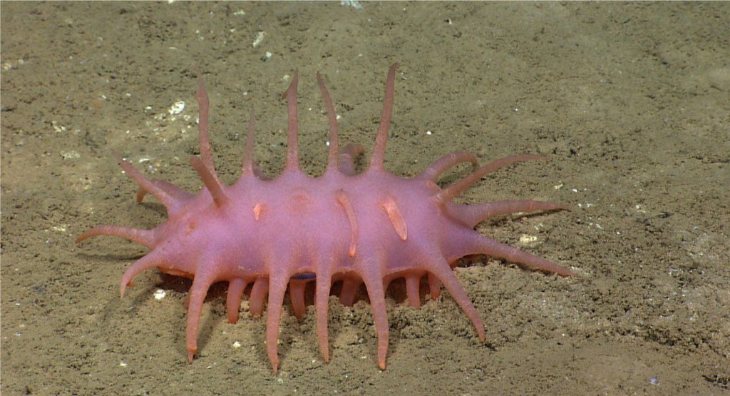
Echinoderms are mostly benthic organisms, living on sandy, rocky, or cavernous reef substrates; some crinoid species can swim. Major groups like the echinoids, asteroids and ophiuroids move around the substrate; sand dollars and heart urchins may reside just below the sediment-water interface. Their domicile water depths range from the shallowest intertidal to abyssal ocean depths – brittle stars and crinoids are frequently found at these great depths. Many species graze algae and organic detritus, some eroding the hard substrates in the process, and other groups like the crinoids and blastoids filter food particles from the water mass. Some species are responsible for the wholesale consumption and decimation of reef coral and bryozoan communities; a well-known example is the voracious, Crown of Thorns starfish on Great Barrier Reef (east Australia).
Echinoderm structure and symmetry
All echinoderms, except the holothurians, have an endoskeleton (below an outer layer of tissue). Shells consist of ossicles – interlocking calcite plates, discs, and spines bound by connective tissue. Many echinoids (particularly sea urchins) possess a spectacular array of rigid spines some of which are poisonous.
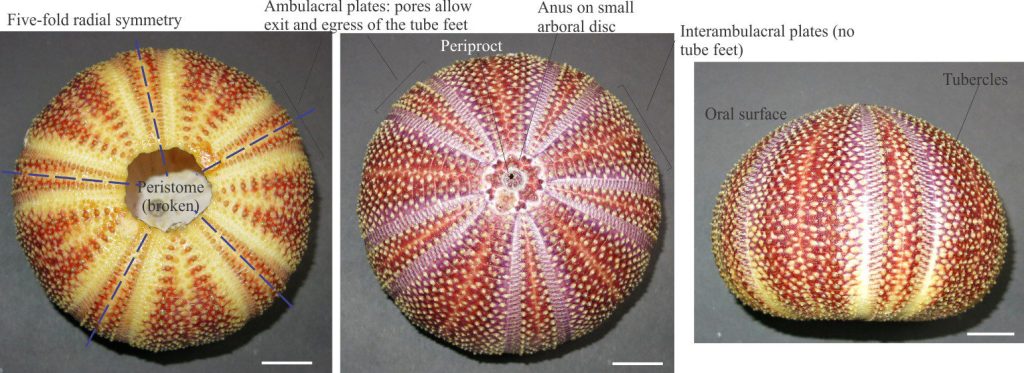
The arrangement of ossicles in all modern and most ancestral echinoderm endoskeletons confers a five-fold, or pentaradial symmetry. In the starfish this is manifested in 5 or more arms (usually in multiples of 5), and in the sea urchins and sand dollars as five-fold radial subdivisions. Crinoid arms are arranged in multiples of 5. Squishy holothurians also have five-fold symmetry, but it is internal and hence not immediately obvious.
Echinoderm orientation
Because of their radial symmetry, there is no ventral-dorsal orientation, or posterior-anterior position on echinoderm tests. Instead, the test is oriented with respect to an oral surface that contains the opening for the mouth, the peristome, and an aboral surface that presents an anal opening – the periproct. On most species the surfaces are on opposite sides of the shell but on some they are on the same side. The oral surface may be down and nearest the substrate (as in the sea urchins, sand dollars, and starfish), or facing upward in species that filter food from the water mass (e.g., crinoids).
The fossil record of echinoderm bits and pieces
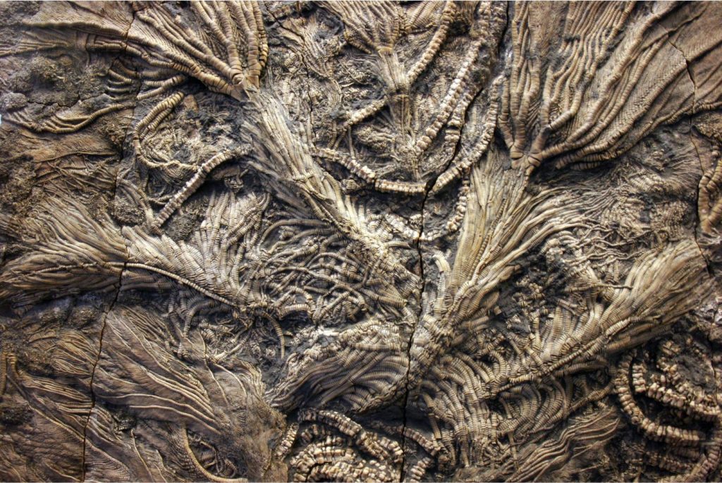
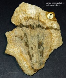
When the animal dies the connective tissue that fixes the ossicles in place decays rapidly. Shells, arms, and columns tend to fall apart, spines disconnect, and thus the fossil record is left mostly with disarticulated specimens although there are some spectacular exceptions (look again at the image at top of the page). Holothurians have microscopic ossicles, but by and large they have very low preservation potential.
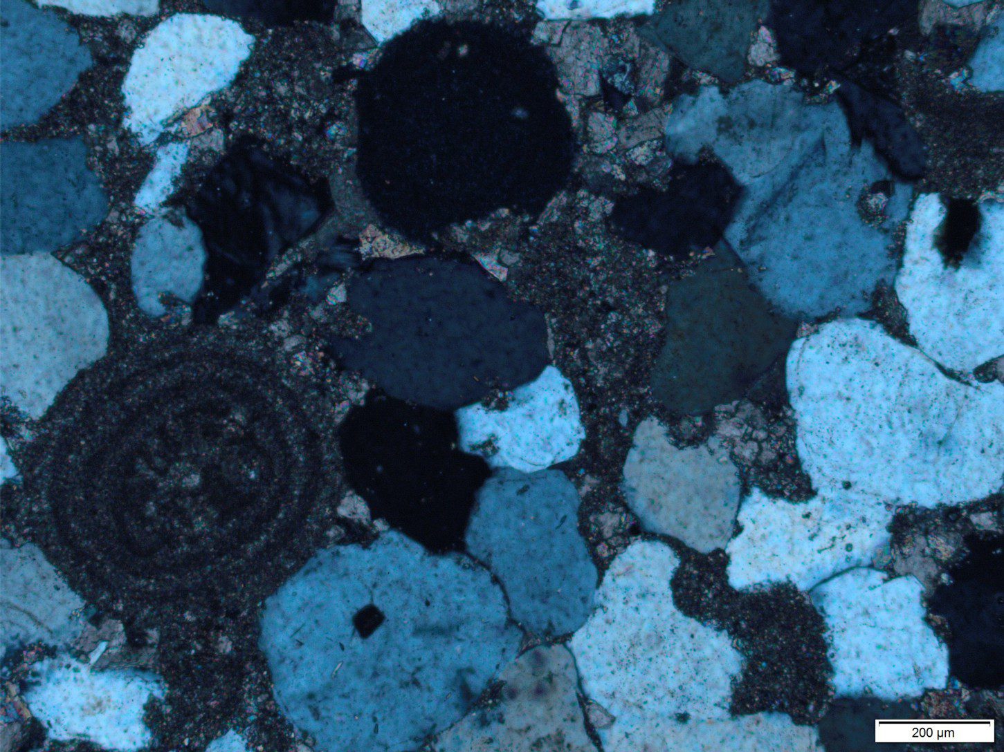
A unique feature of echinoderm plates, discs, and spines is that each fragment consists of a single calcite crystal. In thin section, each will extinguish uniformly under crossed nicols. Furthermore, calcite cements are syntaxial on echinoderm fragments, such that cement and original fragment will extinguish together. Plates and spines are commonly porous, a characteristic that aids their identification.
The diagnostic features of the extant echinoderm classes, that have been dominant since the beginning of the Triassic, are outlined below.
Echinoids
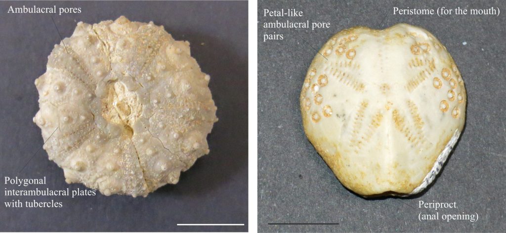
Instead of arms, the sea urchins and sand dollars are divided into five zones, each zone containing ambulacral and interambulacral plates. The ambulacra contain rows of pores that in life provide access to a myriad, bristling tube feet that guide food and water to the mouth along ambulacral grooves – the tube feet are rarely preserved. The interambulacral plates do not contain pores for tube feet but do have tubercles to which spines are attached. In sand dollars, the ambulacral areas are arranged petal-like. Both plates and spines have high preservation potential.
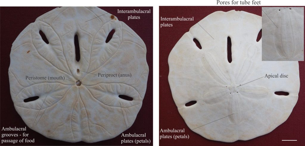
A variation on this theme is found in irregular echinoids, that burrow into the soft substrate. This lifestyle means that the mouth and anus are placed at the margins of the test – the mouth-peristome in front so the animal can feed as it moves through the sediment, and the anus-periproct at the rear (a sensible evolutionary arrangement). Irregular echinoids do not have the same symmetry as ‘regular’ species. An example is shown below.
Asteroids
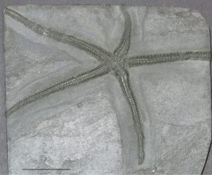
Starfish are flattened versions of sea urchins with the added complication of prominent arms in multiples of five. Tests are made up of ambulacral and interambulacral plates, but these are much smaller than in the echinoids. The oral surface is down; the aboral surface up. Some have spines but they are less prominent than on the sea urchins. All these components are preservable but distinguishing them from echinoids in the fossil record may be difficult.

Right: Star fish using its tube feet, extending from the oral surface (underside), to search for food on the rocky substrate. The outer surface of the arms is lined with small spines. Image credit: NOAA Office of Ocean Exploration and Research, Hohonu Moana 2016
Ophiuroids
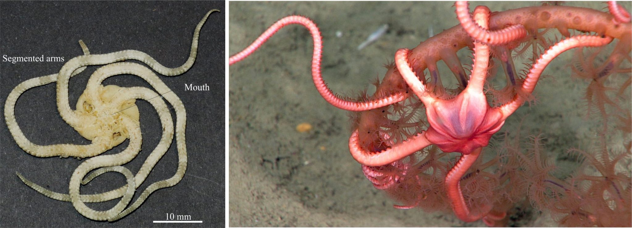
Brittle stars tend to be more delicate than their starfish cousins. They are distinguished by a prominent central disc and body made up of plates, from which the arms extend. The arms are covered by platy ossicles, but unlike the starfish there are hollow tube-like calcite structures through the middle of each arm. The arms are brittle and break easily. The central body may be preserved intact.
Crinoids
Of the two main groups of modern crinoids, the sea lilies are permanently attached to substrates by holdfasts, whereas the feather stars are capable of movement. Fossil crinoids appear to be similar to these two main types. A spectacular example of the stalked variety is shown in the image at the top of the page.
Crinoid tests include plates that make up the main body of the animal (the calyx or arboral cup). The calyx of sea lilies is attached to its root system by a longish, flexible column made up of close-fitted calcite discs, or columnals. The flexible crinoid arms, also constructed of small plates, are attached to the calyx. The arms mostly occur in multiple of five. Flexible, feather-like pinnules that in living forms contain tube feet, extend from the arms; the tube feet guide food particles towards the mouth (the mouth and oral surface in crinoids faces upward).

Right: A sea lily attached by a long stem of columnals to the rocky substrate. The mouth is located at the centre of the central depression from which the arms extend. Extending from the main stalk are cirri that are used for attachment to the substrate. Image credit: NOAA Office of Ocean Exploration and Research, 2017 American Samoa
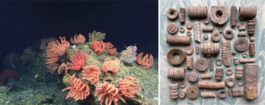
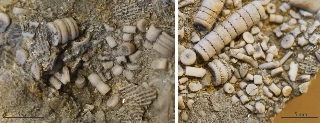
Right is a cornucopia of Carboniferous crinoid columnals, some connected in segments of ancient crinoid stalks. Image courtesy of Dr. Katie Strang on Twitter @palaeokatie and instagram.com/scottishfossils
The most commonly preserved part of a crinoid is its columnals that are easily recognised by their disc-like form, transverse profiles that are round, oval, pentagonal, or star-shaped, all with a central canal (filled with tissue). Like all other echinoderm plate structures, these too consist of a single calcite crystal. Columnals are so common in some limestones that they constitute the primary clast framework. There are also some spectacular examples of complete fossil crinoids.
Sources of information
Several photos were kindly provided by Annette Lokier, University of Derby.
The image of Carboniferous crinoid columnals was kindly provided by Katie Strang Twitter @palaeokatie and instagram.com/scottishfossils
The NOAA Ocean Exploration and Research site has an excellent image library.
The Paleontological Society provides free access to its Digital Atlas of Ancient Life that contains oodles of explanatory texts, field guides, Apps, and images on the fossil record.
Other posts in this series
Bivalve shell morphology for sedimentologists
Gastropod shell morphology for sedimentologists
Brachiopod morphology for sedimentologists
Trilobite morphology for sedimentologists
Cephalopod morphology for sedimentologists
Coral morphology for sedimentologists
Graptolite morphology for sedimentologists
