Geological-logo
Geological-logo

Carbonates in thin section: Forams and sponges

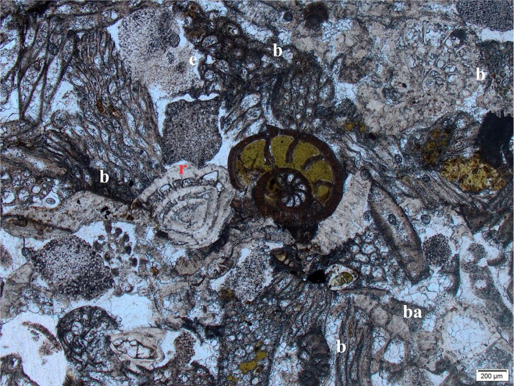
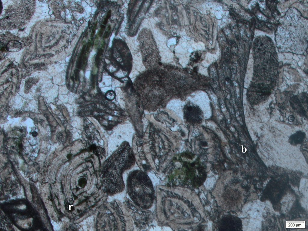
A couple of rotaliids in various states of comminution (possibly Amphistegina – the one labelled ‘r’ is broken along its left margin), and an evolute planispiral foram (centre) in bioclastic grainstone that also contains plenty of bryozoa (b), echinoid plates (e) and spines, and barnacles (ba). The central foram is partly filled with glauconite. Oligocene, New Zealand. Plain polarized light.

Two important contributors to bioclastic limestones – foram tests and skeletal sponge spicules


Foraminifera

Foraminifera (forams) are single celled, marine to brackish water organisms that inhabit the photic zone (their diet consists of algae, diatoms, and other photic zone micro-organisms). They construct exoskeletons (tests) either by secreting calcium carbonate, or by gluing together silt and clay-sized sediment particles (agglutinated forms); there is also a small group of chitinous forms. They are divided into two broad groups: planktic species that spend their lives within the water column, and benthic forms that live on or in the sea floor sediment. A few species encrust other larger shells, corals, and bedrock. Benthic forams are by far the largest and most diverse group.

Forams are sensitive to changes in environmental conditions such as substrate, water depth, salinity, and temperature and thus are excellent paleoenvironmental and paleobathymetric indicators. Their sensitivity to temperature is also reflected in the partitioning of stable oxygen and carbon isotopes, providing excellent proxies for oceanic paleotemperatures. They are also one of the most important fossil groups for high resolution biostratigraphy, particularly in Cretaceous and Cenozoic rocks, a development for which the oil exploration industry can claim much credit.

Forams are microscopic, mostly less than a millimetre across, except for a few species like the iconic Nummulites that in some cases can exceed 50mm diameter. You will need a decent binocular microscope to get any detail out of them; modern foram studies routinely use a scanning electron microscope for species ID.

The variety of test form ranges from single chambered species to those with multiple chambers arranged in linear and flat spiral arrays, and conical spiral forms that look superficially like small gastropods.  Some of the more common forms are shown below.
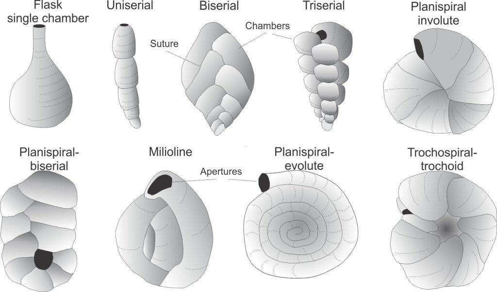
Some of the more common foram test shapes. The original figure is from Loeblich and Tappan, 1964 (the current figure was accessed and modified from https://www.ucl.ac.uk/GeolSci/micropal/foram.html )

Foram classification is based on test morphology, wall structure, and aperture geometry.

The test morphology one sees in thin section depends on the orientation of the fossil. In this situation, the structure of the test wall can be an important guide, at least to the taxonomic level of order.


There are four main orders

Textulariids: Agglutinate  species grow their tests by ‘gluing’ together small sedimentary particles with calcite or silica cement. In thin section they typically appear dark, almost isotropic, although silt-sized quartz, feldspar, and fossil fragments are usually visible.
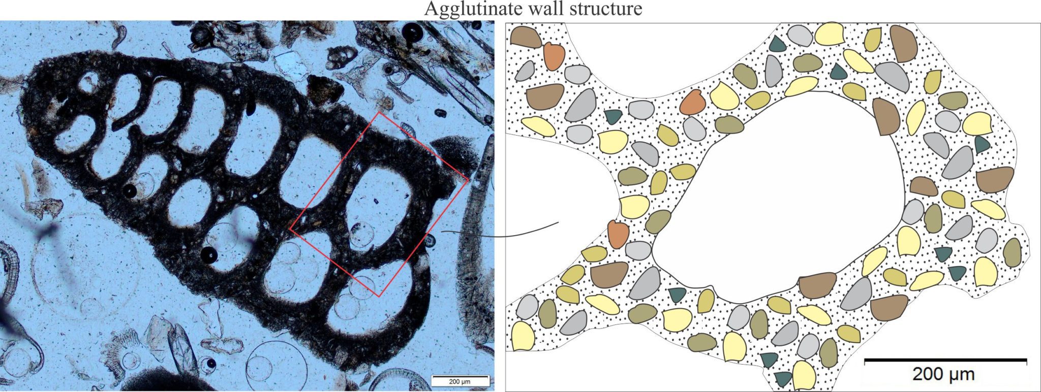
A modern agglutinate foram (Textulariid) from northern New Zealand made up of clay and silt-sized sediment particles (zoom in to see these). Left plain polarized light. Right A schematic representation of a Textulariid chamber and agglutinate particles.

Rotalìids (Hyalina – hyaline means almost transparent). Rotaliids secrete either calcite or aragonite (never a mix of the two) in crystallites arranged in radial fashion with c-axes normal to the test wall. A new layer of radial crystals is precipitated for each chamber addition. Thus, in thin section the foram test walls appear layered. Under crossed polars there may be a pseudo-extinction cross that will rotate with the microscope stage.

This group includes benthic and planktic species, including well-known genera like Nummulites and Globigerina. Test walls have fine perforations (perforate) – these are easy to see in planktic forms. Some species contain microgranular calcite, and a few have walls constructed of large, perforated crystals.

An Oligocene Amphistegina (a Rotaliid) in cross-section showing the characteristic layered and radial, almost prismatic crystal structure. Crystallites are generally normal to the test wall. Plain polarized light. A simplified schematic of this texture is shown on the right. Under crossed polars, crystal extinction appears to sweep across the foram section.


Several Rotaliids, probably Amphistegina, in an Oligocene bryozoa (b)-foram bioclastic grainstone. Chamber walls are layered with crystals arranged in characteristic radial patterns. Cements are mainly drusy calcite. Pale green patches are incipient glauconite cements. Plain polarized light.


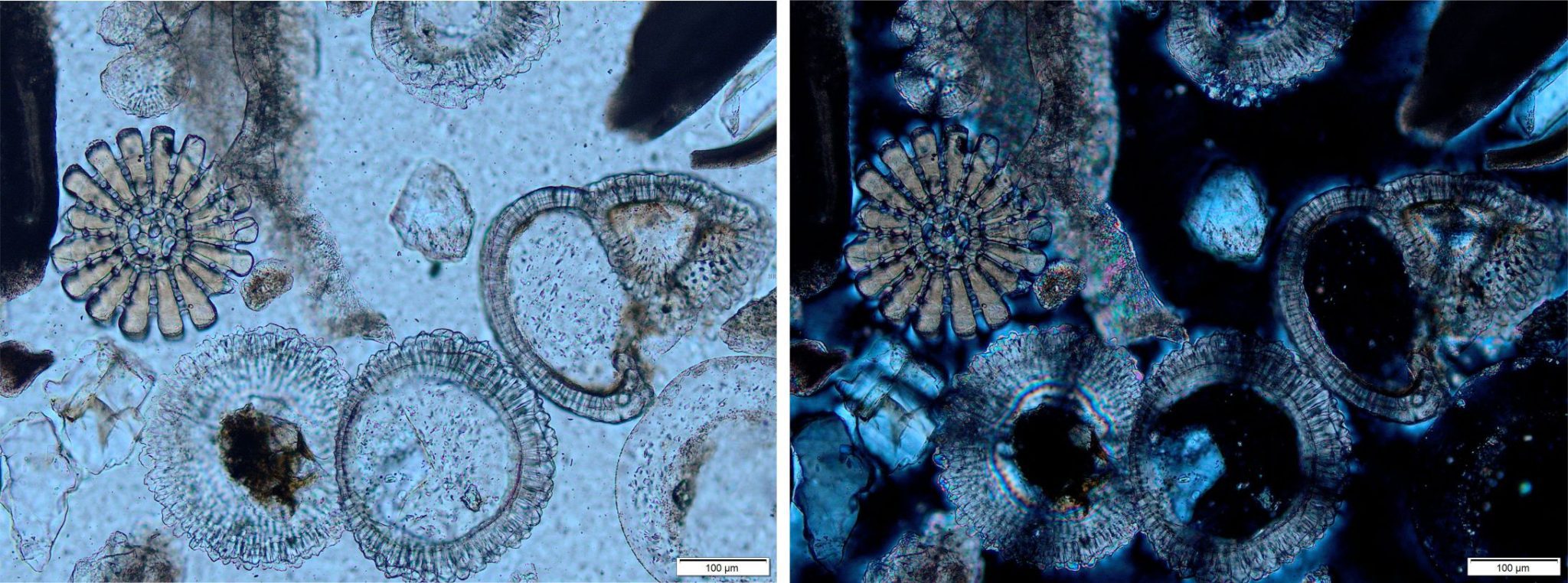
Details of some Recent Rotaliid forams showing the characteristic layered and radial-prismatic crystal textures. Left plain polarized light. Right crossed polars. Note the pseudo-extinction crosses in the forams; each cross will rotate in concert with the microscopic stage. The echinoderm spine cross-section (left) is a single crystal of calcite.


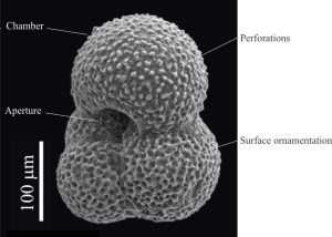
SEM image of the iconic planktic foram, Globigerina, a trochispiral Rotaliid. The test is peppered with micron-sized perforations, or pores. Image credit: Bolli, H. M., Saunders, K. Perch-Nielsen, 1985
SEM image of the iconic planktic foram, Globigerina, a trochospiral Rotaliid. The test is peppered with micron-sized perforations, or pores. Image credit: Bolli, H. M., Saunders, K. Perch-Nielsen, 1985


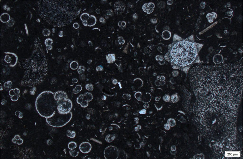
A mudrock with abundant planktic forams. Zoom in on the large foram lower left to see the radially arranged wall crystallites and spinose ornamentation. Most of the forams are intact, indicating relatively gentle suspension deposition from the overlying water column. There is a single fragment of calcareous algae bottom right. The star-shaped fossil (top right) looks similar to modern star-shaped forams (Baclogypsina sphaerulata) found on some Indo-Pacific beaches, particularly Okinawa where they are known as star sands. Sample source and age unknown.


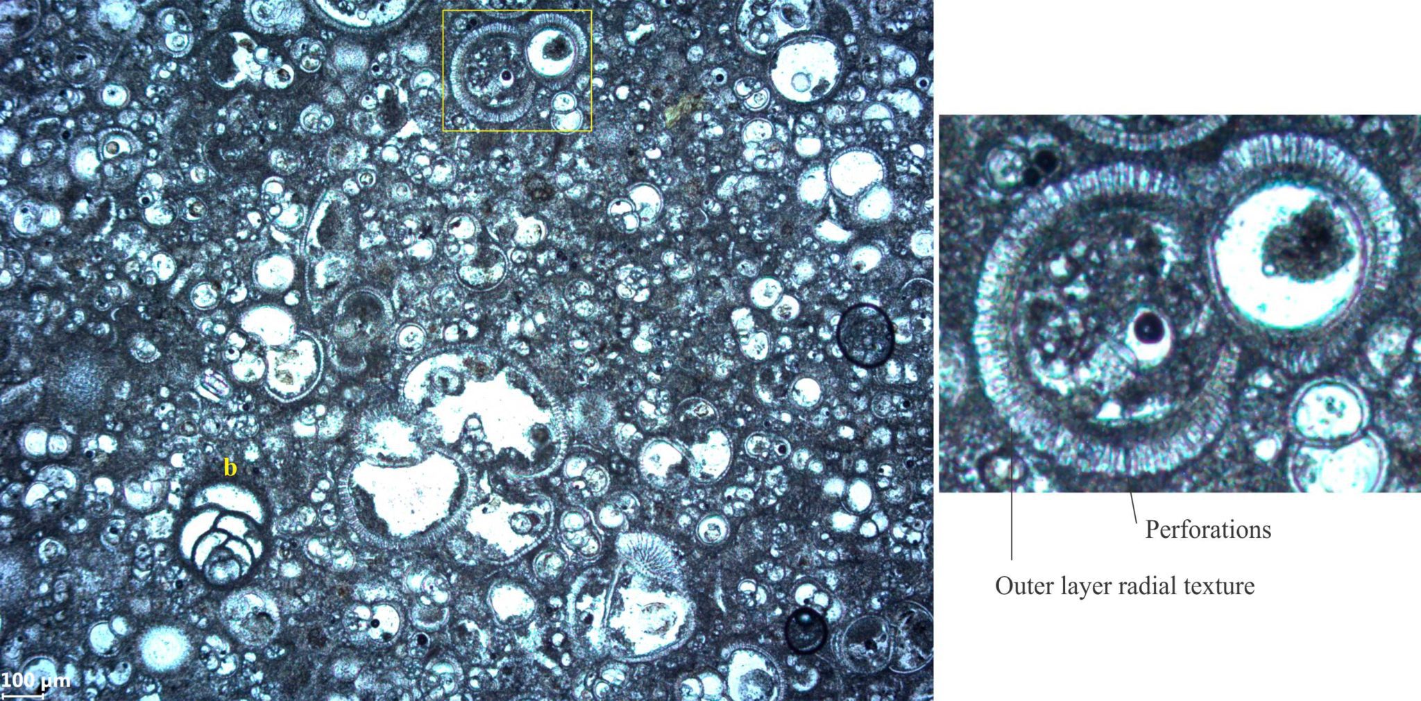
Foraminifera ooze containing abundant planktic rotaliid forms (Globigerina-like), plus a few miliolid forams (microgranular texture – see description below) – note the biserial form near bottom left (b). The rotaliids show typical radial crystal walls with perforations extending the entire wall thickness, and spinose ornamentation, shown in greater detail in the inset image. Most chambers are filled by coarse crystalline calcite. Image courtesy of Mark Lawrence, GNS.


Fusulinids are an order that died off during the end Permian mass extinction. The walls of this group consist of microscopic, equigranular calcite. Many, but not all, are perforate.

Left: Bedding of Permian grainstone exposing a Fusulinid multitude; the elongate forams have been aligned by marine currents. Right is a schematic of typical fusuline microgranular calcite wall structure. The c-axes of individual calcite crystals are oriented randomly – under crossed polars the crystallites will also extinguish randomly as the microscope stage is rotated. The example is from South Bay, Ellesmere Island.


Miliolids Porcellanous (smooth and commonly shiny) – This group has walls constructed of high-magnesium, acicular to equant calcite crystals, generally lacking preferred orientation. Tests are imperforate (cf. the Fusulinids).

A biserial foram with Miliolid, or porcellaneous wall structure (left), consisting of needle and/or equant calcite crystallites that have no preferred orientation. Right is a schematic representation of this wall structure.


Sponges

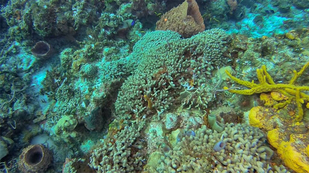
A nice collection of extant sponges living amongst Porites corals in Columbia reef, Cozumel. On the right, yellow tube and entrusting species; lower centre-Left pale blue vase sponges; and lower left and top, three large barrel sponges.
A nice collection of extant sponges living amongst Porites corals in Columbia reef, Cozumel. On the right, yellow tube and encrusting species; lower centre-left pale blue vase sponges; and lower left and top, three large barrel sponges. Image courtesy of Charlie Kerans.

Sponges (phylum Porifera) are the simplest of multicell invertebrates. They survived multiple extinction events since the Cambrian; there are hints that they may have evolved during the late Precambrian. Most forms have an internal scaffolding-like skeleton made up of solid rod-like structures called spicules (or sclerites), that are bound together to form a semi-rigid framework, around which the spongin grows and is structurally supported (spongin, as in a bath sponge, is an insoluble protein). The spicules are composed of amorphous silica (opal-like), or high-magnesium calcite – the former are more common. Siliceous sponges are also called glass sponges. Spicules also focus light in much the same way as optic fibres – an interesting discovery. The spicules disaggregate when the animal dies. Sponge taxonomy is based primarily on spicule geometry.

Sponge spicules come in many shapes, usually less than a few millimetres long. They occur as single rays, pointy at the growing end, or as complex architectures that radiate from a central point. Each species has a unique spicule size and shape. Calcareous spicules (Calcarea) are single crystals. Siliceous spicules have a central canal.

All spicules are prone to recrystallization during burial diagenesis, partly because of their composition (the amorphous silica will recrystallize to quartz), but also because of their relatively high surface area. Siliceous spicules may also be replaced by carbonate and in the process provide an important source of dissolved silica to ambient geofluids.
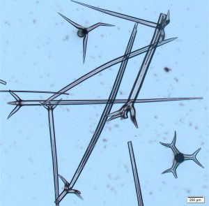
 

Disaggregated siliceous sponge spicules at different orientations in Recent sediment, northernmost New Zealand. The longest is about 2.2mm. The spicules in this group are called Tetraxons having three rays of equal length plus a much longer ray, all radiating from a single point. Variations on this theme include spicules with one, three, and six or more rays. The central canals appear as dark lines within each ray.


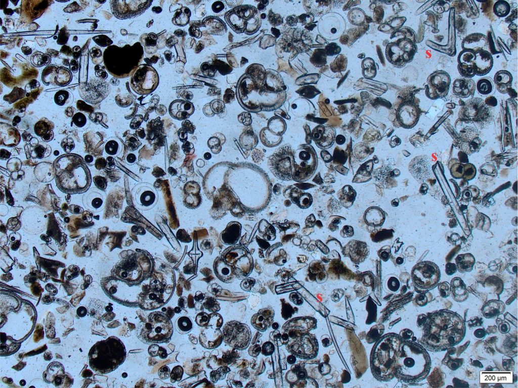
A grain-mount thin section of Recent bioclastic sediment from northern New Zealand. There are abundant Rotaliid forams (radial wall structure) plus a variety of intact and broken sponge spicules (s) that present as clear, colourless, pin-like rods. Plain polarized light.


Acknowledgement

Many thanks to Kirsty Vincent, Earth Sciences, Waikato University for access to the petrographic microscope.


Other links

https://foraminifera.eu/about.html Lots of info and images on forams

R.G.C. Bathurst, 1976. Carbonate Sediments and their Diagenesis. Always worth a read

SEPM STRATA has a good section on forams


Other posts in this series

Brachiopod morphology for sedimentologists

Bivalve shell morphology for sedimentologists

Gastropod shell morphology for sedimentologists

Cephalopod morphology for sedimentologists

Optical mineralogy: Some terminology

Carbonates in thin section: Bryozoans

Carbonates in thin section: Echinoderms and barnacles

Carbonates in thin section: Molluscan bioclasts

Neomorphic textures in thin section

Sandstones in thin section

Greywackes in thin section

Mineralogy of carbonates; skeletal grains

Bivalve morphology for sedimentologists

Mineralogy of carbonates; non-skeletal grains

Mineralogy of carbonates; lime mud

Mineralogy of carbonates; classification

Mineralogy of carbonates; carbonate factories

Mineralogy of carbonates; basic geochemistry

Mineralogy of carbonates; cements

Mineralogy of carbonates; sea floor diagenesis

Mineralogy of carbonates; Beachrock

Mineralogy of carbonates; deep sea diagenesis

Mineralogy of carbonates; meteoric hydrogeology

Mineralogy of carbonates; Karst

Mineralogy of carbonates; Burial diagenesis

Mineralogy of carbonates; Neomorphism
Mineralogy of carbonates; Pressure solution

Mineralogy of carbonates: Stromatolite reefs

Leave a Comment

Your email address will not be published. Required fields are marked *

Archives
Categories
Recent Posts
Scroll to Top