Some basic bivalve morphology to assist sedimentological interpretations
Ok, you’re not a paleontologist but during a sedimentological – stratigraphical examination of outcrop or core you encounter body fossils – whole and fragmented shells, carapaces, skeletons, or rigid frameworks, as scattered occurrences, or packed communities. Notwithstanding their potential biostratigraphic value, identification of these fossil critters will provide important clues about depositional processes and paleoenvironments, paleoecology, unconformities and condensed sections, and taphonomy (how dead critters are preserved). The circumstances of field work may mean that your eager paleontologist colleague cannot stand at the outcrop and regale you with all the observable taxonomic and paleoecological information you need. That task is appointed to you.
Identifying fragments even to the level of phylum is a useful beginning; are they molluscs or brachiopods, corals or bryozoa. It is a truism that the larger or more complete a fossil, the easier it will be to identify to any taxonomic level. However, even with small fragments there may be distinguishing features that allow identification to Class or Order levels of taxonomy. For example, it will frequently be possible to determine whether fragments of molluscs are bivalves (pelecypods), gastropods, or ammonites?
Here is a brief outline of some important features of bivalve morphology that may help your identification of fossil forms. The focus is on external and internal shell morphology and how to orient your specimens for correct description. How you describe and photograph these fossils will determine the confidence you and your associates have for any future interpretation.
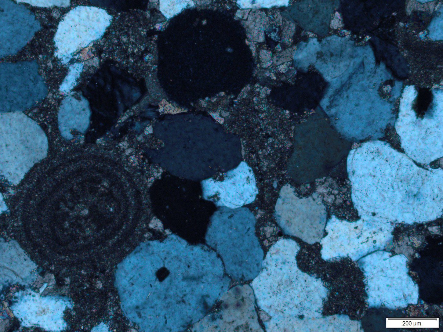
Identification of bivalves in thin section will also provide valuable information.
Other sources
There are lots… but here are a couple of links to recent texts.
The Paleontological Society provides free access to its Digital Atlas of Ancient Life that contains oodles of explanatory texts, field guides, Apps, and images on the fossil record.
Bringing Fossils to Life: An Introduction to Paleobiology, Donald Prothero. Now in its 3rd Edition.
Shell orientation
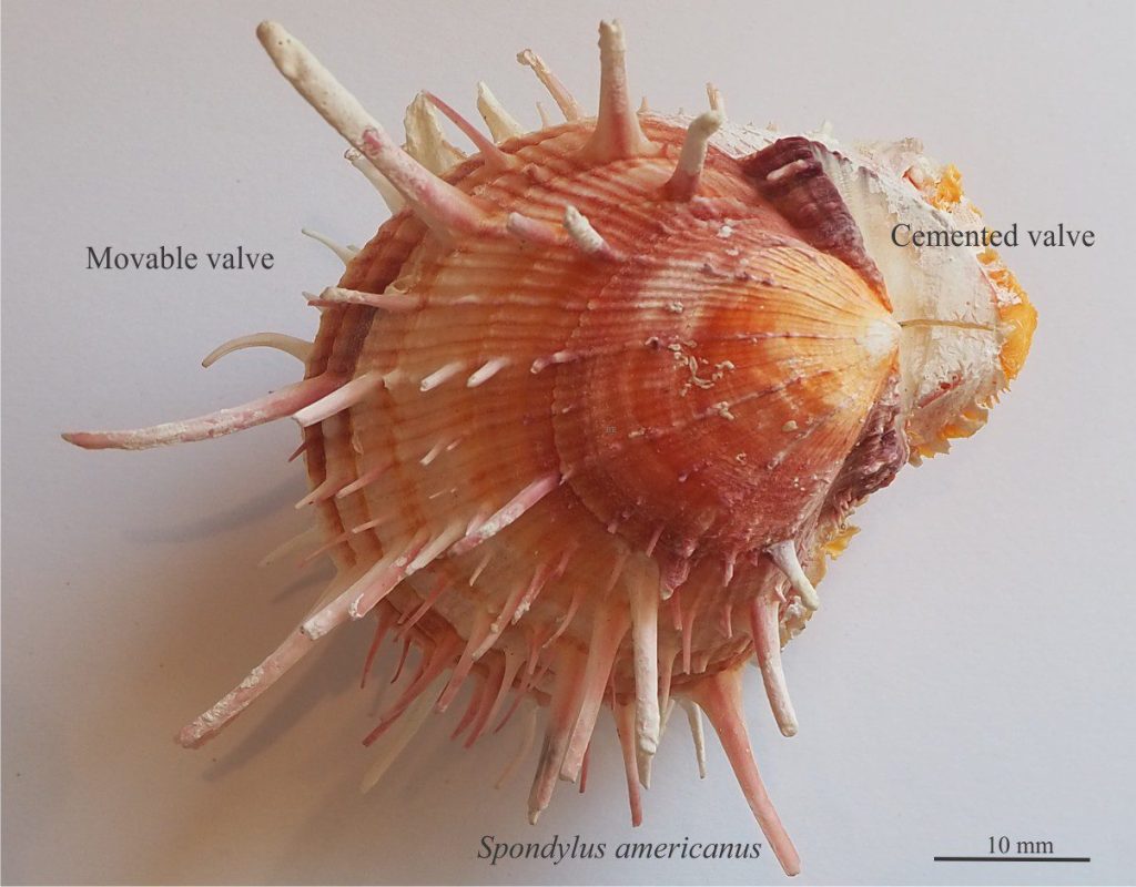
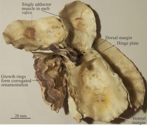
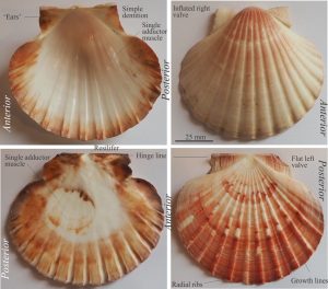
The most common bivalve configuration is two equal valves that are bilaterally symmetrical, which means when you view an articulated shell edge-on, the two valves are mirror images. There are lots of exceptions, the most common where one valve is cemented to a substrate (usually the larger valve), as is usually the case for oysters (e.g., Ostrea). Free swimmers like scallops (e.g., Pecten) are also inequivalve.
[Brachiopods are also bivalved; the valves are inequal but are equilateral such that a plane of symmetry lies along a line through their centres.]
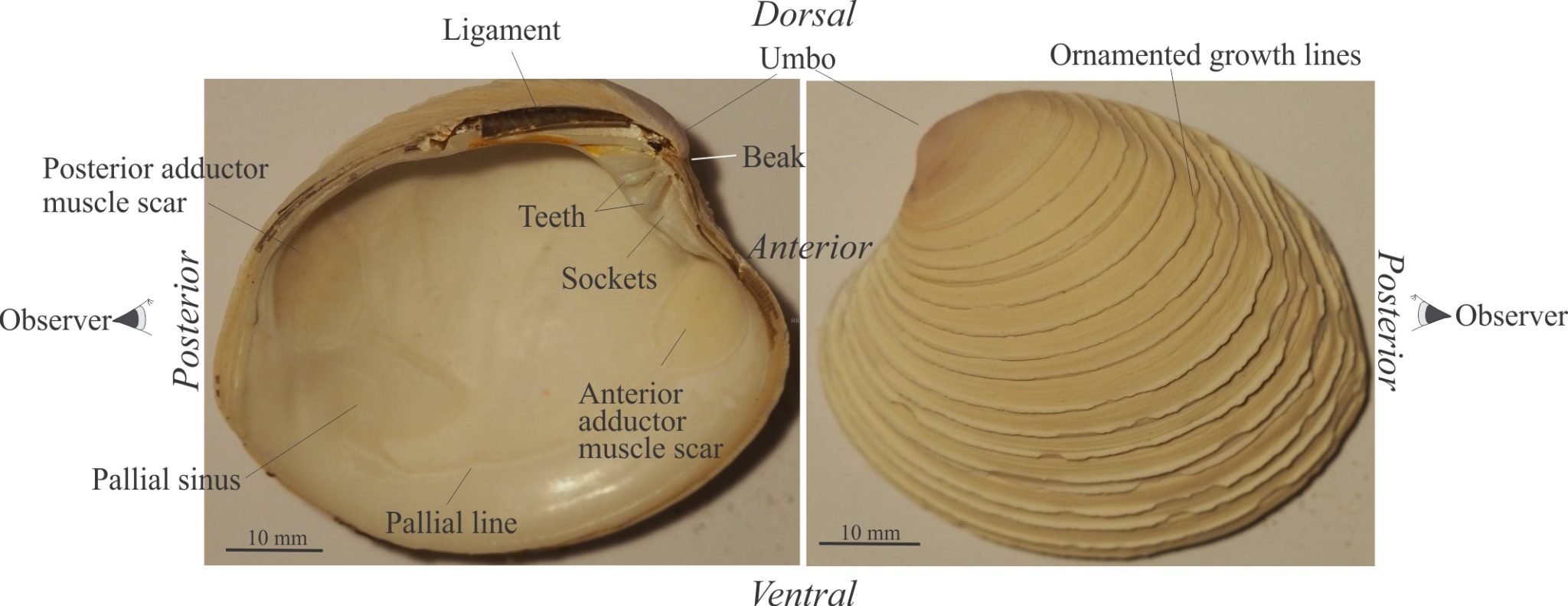
Left valve – right valve: The standard procedure for determining valve orientation is to hold the shell upright with the beak pointing away from you. The valve on the left is left valve. The shell margin closest to you is posterior; away from you is anterior. Identification of left-right valves in cemented bivalves is more complicated and may require knowledge of the animal’s anatomy. Some modern oysters like Ostrea cement their right valve (the left valve acts as a smaller lid), whereas other species cement the left valve.
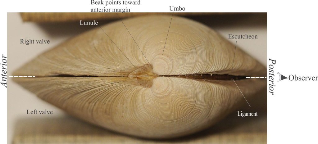
Dorsal – ventral: The part of the shell containing the umbo, beak and hinge-ligament structures is dorsal; the opposite, outer shell margin is ventral. In articulated specimens the ventral margin may be closed (as in the two examples shown above), or open as in the common Razor Clam.
External bivalve morphology
Beak: The pointy end of the shell (always dorsal) that represents the initial stage of animal growth. It points towards the anterior margin.
Escutcheon: A narrow, oval-shaped depression along the dorsal margin of both valves. It is posterior to the beak and approximately parallels the hinge line and the ligament.
Growth rings/lines: Concentric bands or ridges that represent changes in growth and calcium carbonate secretion; note the commonly held notion that each ring represents annual or seasonal patterns is not supported by detailed studies of bivalve ages. The rings are centred about the umbo. Depending on the species, rings may be delicate lines, low-relief ridges, or contain more complicated ornamentation, such as the platy growths on the example of Bassina shown above. If radial ribs are present, their intersection with growth rings may produce more ornate structures such as tubercles or spines.
Lunule: A small heart-shaped depression immediately below and anterior to the beak.
Ornamentation: The most common type presents raised ribs that radiate from the beak towards the ventral margins. Like growth rings, they can assume several different geometries, from narrow, low-relief ridges to more prominent corrugations like those commonly seen in Pectens (scallops). The intersection of growth rings and radial ribs in some species can produce in some spectacular nodular or spiny growths. An excellent example of the latter is seen in Spondylus americanus (shown at top of page).
Umbo: The dorsal part of a valve where curvature increases significantly; this region also contains the beak.
Internal bivalve morphology
Adductor muscle scars: Variably shaped scars where the animal’s adductor muscles are attached to the shell. Two muscle scars are present in most equivalve species near the anterior and posterior margins of both valves. A single scar is present in some inequivalve species such as oysters and scallops.
Bivalve shells are open and closed by their adductor muscles. When the valves are closed a chitinous ligament, that help keep the valves attached along a hinge line, is under tension (it acts a bit like a rubber band). When the muscles relax, the ligament, is no longer under tension; it relaxes, and the valves open so that the animal can do its thing. This is the reason that most fossil bivalves are preserved with valves open. [the opposite condition exists in brachiopods].
Dentition: The locus of bivalve articulation is the hinge line along which is an array of teeth and sockets. The teeth and sockets on one valve have corresponding sockets and teeth on the other valve. There is great variety in the number, size, and shape of these dentition elements, but the two most common types are heterodont and taxodont dentition. Examples are shown below.
Heterodont dentition consists of two or three largish cardinal teeth and corresponding sockets that grow immediately below the beak. In many species (but certainly not all) there are lateral teeth that extend along the hinge line towards the anterior and posterior margins.
Taxodont dentition consists of many small teeth and sockets arranged in a row, usually on either side of the beak.
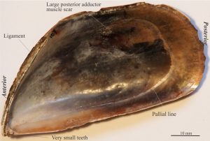
Heterodont dentition is the most common type but there are several variations. For example, dentition in mussels consists of two or three very small teeth below a beak that is strongly skewed towards the anterior margin – this is referred to as dysodont dentition. Dentition in the common scallop consists of a few narrow, low relief ridges along a straight hinge line – this is a type of isodont dentition.
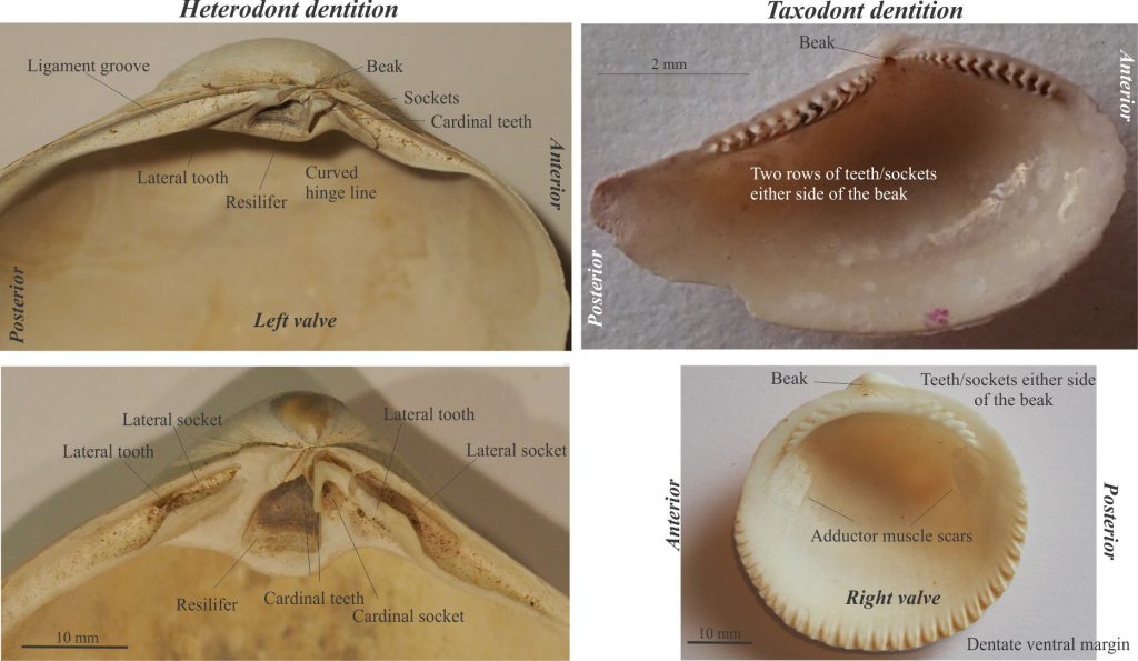
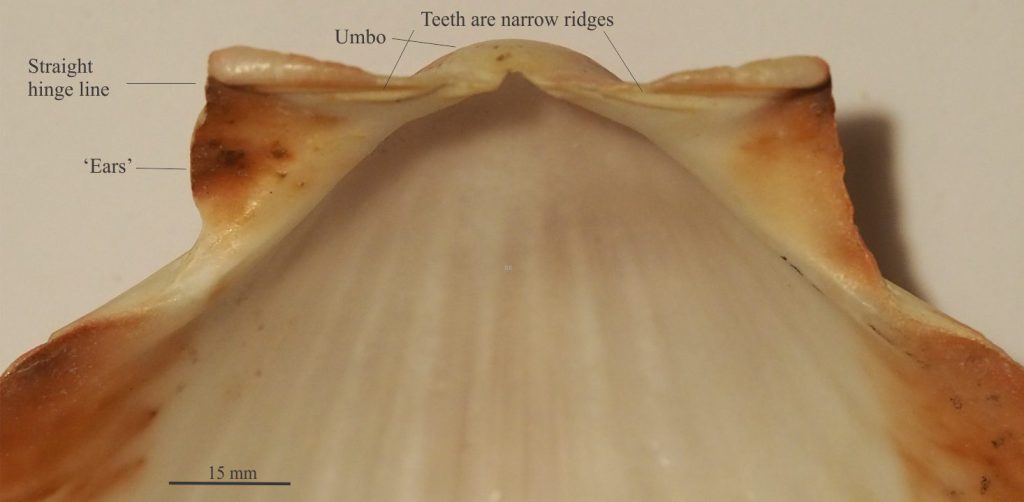
Hinge line: Most bivalves are hinged by teeth and sockets along a curved or straight line located below the beak. If a resilifer pit is present, then the hinge line is divided into anterior and posterior segments.
Ligament: A tough, elastic, complex protein (called conchiolin) secreted by the animal, that helps keep the two valves attached, and assists in articulation. When the valves are closed the ligament is under tension; when open the ligament is relaxed. The ligament itself has very low preservation potential but the groove in which it sits may be preserved.
Pallial line and pallial sinus: (pallium is Latin for mantle or cloak). The pallial line marks the edge of the mantle that in life covers the animal viscera. It is commonly etched into the shell and occurs on both valves.
Bivalves that possess siphons commonly have a pallial sinus, an indentation in the pallial line that marks the position of the siphons (siphons are used for feeding, respiration, and locomotion). It is always posterior. It too is commonly etched into the shell and may be preserved in fossil specimens.
Resilifer pit: A roughly triangular-shaped depression just below the beak and present in both valves. In life it is occupied by fibrous, proteinous conchiolin that assists in valve articulation.
Other posts in this series
Echinoderm morphology for sedimentologists
Trilobite morphology for sedimentologists
Brachiopod morphology for sedimentologists
Cephalopod morphology for sedimentologists
Gastropod shell morphology for sedimentologists
Carbonates in thin section: Molluscan bioclasts
Mineralogy of carbonates; skeletal grains
Mineralogy of carbonates; non-skeletal grains
Mineralogy of carbonates; lime mud
Mineralogy of carbonates; classification
Mineralogy of carbonates; carbonate factories
Mineralogy of carbonates; basic geochemistry
Mineralogy of carbonates; cements
Mineralogy of carbonates; sea floor diagenesis
Mineralogy of carbonates; Beachrock
Mineralogy of carbonates; deep sea diagenesis
Mineralogy of carbonates; meteoric hydrogeology
Mineralogy of carbonates; Karst
Mineralogy of carbonates; Burial diagenesis
Mineralogy of carbonates; Neomorphism
